Introduction
In the realm of pathology, peripheral blood smear image analysis stands as a critical component of routine laboratory diagnostics. However, manual examination of these images can be labor-intensive, time-consuming, and prone to interobserver variability. This challenge has spurred significant research into developing automated image analysis systems, with a focus on enhancing diagnostic accuracy and efficiency.
Key Steps in Image Analysis
The process of peripheral blood smear image analysis involves several key steps:
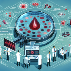
- Image Segmentation: This initial step involves isolating the object or region of interest from the complex background of the blood smear image. Techniques such as Support Vector Machine (SVM) and Artificial Neural Networks (ANNs) are commonly employed.
- Feature Extraction and Selection: The goal here is to derive distinctive characteristics from the segmented objects, facilitating the classification process. Effective feature selection is crucial to minimize overlap between classes and enhance classification accuracy.
- Pattern Classification: This final step involves assigning a class to the selected features using supervised or unsupervised learning algorithms. Combining multiple classifiers can further improve discrimination.
Enhancing Practitioner Skills
Practitioners can significantly enhance their diagnostic capabilities by integrating these advanced image analysis techniques into their workflow. Here are some actionable steps to consider:
- Adopt Machine Learning Tools: Utilize machine learning algorithms such as SVM and ANN for more accurate and efficient image segmentation and classification.
- Engage in Continuous Learning: Stay updated with the latest research and advancements in image analysis to continually refine diagnostic skills.
- Collaborate with Experts: Work alongside data scientists and engineers to develop customized algorithms tailored to specific diagnostic needs.
Future Directions
As the field of peripheral blood smear image analysis continues to evolve, future research should focus on developing software tools that integrate the best-performing algorithms with data mining techniques. This integration will automate and enhance the accuracy of blood smear image analysis, ultimately improving patient outcomes.
Conclusion
The journey towards automating peripheral blood smear image analysis is a promising one, with significant potential to transform routine laboratory tasks into precise, efficient processes. By embracing these advancements, practitioners can enhance their diagnostic accuracy and contribute to better healthcare outcomes.
To read the original research paper, please follow this link: Peripheral blood smear image analysis: A comprehensive review.


