Introduction
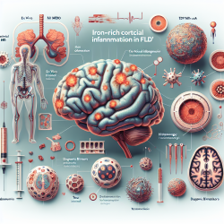
Frontotemporal lobar degeneration (FTLD) is a complex spectrum of neurodegenerative diseases characterized by two main pathologic categories: tau (FTLD-Tau) and TDP-43 (FTLD-TDP) proteinopathies. These conditions often present similar clinical symptoms, complicating accurate diagnosis and treatment. Recent research by Tisdall et al. (2021) has unveiled novel insights into the iron-rich cortical inflammation that distinguishes these subtypes, offering new avenues for diagnosis and therapy.
Research Highlights
The study employed an integrated approach using ex vivo MRI and histopathology to explore the distinct iron-rich pathologies in FTLD. Key findings include:
- Identification of novel iron-rich inflammation patterns in FTLD-Tau and FTLD-TDP.
- FTLD-TDP showed prominent superficial cortical layer iron reactivity, while FTLD-Tau exhibited deep cortical layer inflammation.
- Iron-sensitive T2*-weighted MRI could potentially serve as an in vivo biomarker for distinguishing between FTLD proteinopathies.
Implications for Practitioners
For practitioners, these findings underscore the importance of advanced imaging techniques in the differential diagnosis of neurodegenerative diseases. Implementing T2*-weighted MRI in clinical settings could enhance the accuracy of FTLD diagnosis, allowing for more tailored therapeutic interventions. Furthermore, understanding the distinct inflammatory patterns can guide the development of targeted therapies aimed at mitigating the specific pathologic processes of each FTLD subtype.
Encouraging Further Research
While this study provides significant insights, it also opens the door for further research. Future studies could focus on adapting these imaging techniques for in vivo use, potentially revolutionizing how FTLD is diagnosed and managed. Additionally, exploring the underlying mechanisms of iron-rich inflammation could lead to novel therapeutic targets, offering hope for more effective treatments.
Conclusion
The research by Tisdall et al. marks a pivotal step in understanding FTLD's complex pathology. By leveraging advanced imaging and histopathological techniques, practitioners can enhance diagnostic accuracy and develop more effective treatment strategies. As we continue to unravel the intricacies of neurodegenerative diseases, such innovative approaches will be crucial in improving patient outcomes.
To read the original research paper, please follow this link: Ex vivo MRI and histopathology detect novel iron-rich cortical inflammation in frontotemporal lobar degeneration with tau versus TDP-43 pathology.