Introduction
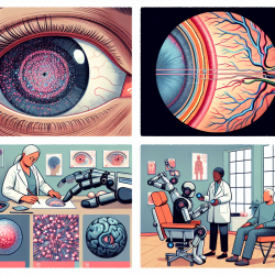
The realm of special education and therapy services is constantly evolving, with new research and technologies emerging to enhance the quality of care provided to students with special needs. One such advancement is the use of automated retinal layer segmentation in CLN2-associated disease, as detailed in the research article "Automated Retinal Layer Segmentation in CLN2-Associated Disease: Commercially Available Software Characterizing a Progressive Maculopathy." This study offers valuable insights into the potential of automated segmentation software to monitor retinal degeneration in patients with CLN2 disease, a rare but significant neurodegenerative disorder.
Understanding CLN2-Associated Disease
CLN2-associated disease, also known as late-infantile neuronal ceroid lipofuscinosis, is a hereditary disorder characterized by progressive brain and retinal deterioration. The disease typically manifests between the ages of 2 and 4, leading to symptoms such as speech delay, motor decline, seizures, and vision loss. The research highlights the importance of early detection and monitoring of retinal changes using optical coherence tomography (OCT) and automated segmentation software.
Key Findings and Implications for Practitioners
The study reveals that parafoveal outer nuclear layer (ONL) thinning is an early indicator of retinal degeneration in CLN2 disease, occurring before significant changes in other retinal layers. This finding underscores the potential of automated segmentation software to provide sensitive, quantitative biomarkers for assessing retinal degeneration. For practitioners, this means:
- Enhanced Monitoring: Automated segmentation allows for precise monitoring of retinal changes, enabling early intervention and better management of CLN2-associated retinal degeneration.
- Objective Assessment: The software provides objective, repeatable measurements, reducing reliance on subjective assessments and improving diagnostic accuracy.
- Informed Decision-Making: By understanding the progression of retinal degeneration, practitioners can make informed decisions regarding therapeutic interventions and patient care.
Encouraging Further Research
While the study provides significant insights, it also highlights the need for further research to validate the findings and explore additional applications of automated segmentation software. Practitioners are encouraged to engage in research initiatives, collaborate with academic institutions, and contribute to the growing body of knowledge in this field. By doing so, they can help refine diagnostic tools and improve outcomes for patients with CLN2 disease.
Conclusion
The integration of automated retinal layer segmentation into clinical practice represents a promising advancement in the management of CLN2-associated disease. By leveraging this technology, practitioners can enhance their diagnostic capabilities, provide more effective care, and contribute to the ongoing development of innovative therapeutic strategies. To delve deeper into the research findings, practitioners can access the original research paper by following this link: Automated Retinal Layer Segmentation in CLN2-Associated Disease: Commercially Available Software Characterizing a Progressive Maculopathy.


