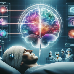
An awake craniotomy is a complex yet invaluable surgical procedure that allows for the preservation of critical brain functions while removing tumors. This technique is particularly beneficial for patients with gliomas near eloquent brain areas responsible for language and speech. However, the success of such surgeries heavily relies on the careful selection and titration of anesthetic agents, especially in patients with pre-existing language deficits.
The Case Study: A Deep Dive into Anesthetic Management
The research article titled "Anesthetic Selection for an Awake Craniotomy for a Glioma With Wernicke’s Aphasia: A Case Report" provides an insightful case study that highlights the importance of meticulous anesthetic management. The case involved a 73-year-old woman with a left temporal lobe glioma near Wernicke’s area, presenting significant language deficits.
The surgical team employed an asleep-awake-asleep (SAS) approach using intraoperative neuronavigation, 5-aminolevulinic acid fluorescence, and awake speech mapping. This method required precise anesthetic titration to optimize intraoperative language testing while ensuring patient comfort and safety.
Key Takeaways for Practitioners
- Patient Selection: Carefully assess patients' physical and psychological readiness for awake craniotomy. Those with severe language deficits or high anxiety may not be suitable candidates.
- Anesthetic Titration: Use the minimum effective dose of sedatives to maintain patient comfort without compromising language testing capabilities.
- BIS Monitoring: Employ bispectral index (BIS) monitoring to guide anesthetic depth and avoid excessive sedation that could hinder language mapping.
- Drug Selection: Consider using dexmedetomidine for its respiratory-preserving properties and propofol for its ability to reduce intraoperative seizures.
The Role of BIS Monitoring
BIS monitoring played a crucial role in this case by helping maintain adequate anesthesia depth without over-sedation. This tool can be particularly useful in preventing patient movement during critical phases of surgery and ensuring active participation in language mapping tasks.
Encouraging Further Research
This case study underscores the need for further research into the effects of different anesthetic agents on intraoperative language mapping. As no single agent has been identified as superior, practitioners are encouraged to stay informed through ongoing studies and adapt their practices accordingly.
Conclusion
The successful outcome of this case highlights the importance of tailored anesthetic strategies in awake craniotomies. By carefully selecting and titrating anesthetics, practitioners can significantly improve surgical outcomes and preserve vital brain functions in patients with gliomas affecting language areas.
To read the original research paper, please follow this link: Anesthetic Selection for an Awake Craniotomy for a Glioma With Wernicke’s Aphasia: A Case Report