Introduction
In the ever-evolving field of neurodegenerative disorder diagnosis, the integration of advanced imaging techniques has proven transformative. The recent study titled "Additive value of [18F]PI-2620 perfusion imaging in progressive supranuclear palsy and corticobasal syndrome" provides compelling evidence of how imaging can enhance diagnostic accuracy and patient outcomes. This blog aims to distill the key findings of this study, encouraging practitioners to harness these insights to improve their diagnostic acumen and patient care strategies.
The Study at a Glance
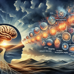
The research investigated the efficacy of early-phase [18F]PI-2620 PET imaging as a biomarker for neurodegenerative diseases, specifically focusing on four-repeat tauopathies (4RTs) like progressive supranuclear palsy (PSP) and corticobasal syndrome (CBS). The study encompassed 78 patients with 4RTs, 79 with other neurodegenerative diseases, and 12 age-matched controls, offering a robust dataset for analysis.
Key Findings
- Regional Hypoperfusion: The study found significant hypoperfusion in 21 out of 246 brain regions, particularly in the thalamus, caudate nucleus, and anterior cingulate cortex. This mirrors the known topology of neuronal injury in 4RTs.
- Discriminatory Power: The perfusion pattern expression showed promise in distinguishing 4RTs from other neurodegenerative disorders, with an area under the curve (AUC) of 0.850. This was further enhanced to 0.903 when combined with tau pattern expression.
- Correlation with Clinical Severity: Perfusion patterns were more closely associated with clinical severity (PSP rating scale and activities of daily living) than tau pattern expression alone.
Implications for Practitioners
For speech-language pathologists and other practitioners, these findings underscore the importance of integrating advanced imaging techniques into diagnostic protocols. The ability to identify specific perfusion patterns can significantly enhance the accuracy of diagnosing 4RTs, leading to more tailored and effective treatment plans.
Moreover, understanding the correlation between perfusion patterns and clinical severity can aid in monitoring disease progression and adjusting therapeutic interventions accordingly. This approach not only improves patient outcomes but also aligns with data-driven decision-making, a cornerstone of modern healthcare.
Encouraging Further Research
While the study provides a solid foundation, it also opens avenues for further research. Practitioners are encouraged to explore the potential of combining perfusion and tau imaging in other neurodegenerative disorders, assessing its utility in different clinical settings and patient populations.
Additionally, longitudinal studies could provide deeper insights into how these imaging biomarkers correlate with disease progression over time, offering valuable information for developing new therapeutic strategies.
Conclusion
The integration of [18F]PI-2620 perfusion imaging into clinical practice represents a significant leap forward in diagnosing and managing neurodegenerative disorders. By embracing these advancements, practitioners can enhance their diagnostic capabilities, improve patient outcomes, and contribute to the broader field of neurodegenerative research.
To read the original research paper, please follow this link: Additive value of [18F]PI-2620 perfusion imaging in progressive supranuclear palsy and corticobasal syndrome.