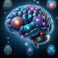
The brain is a dynamic organ that continuously evolves in response to genetic and environmental factors. This evolution is not just limited to function but extends to its very structure. Thanks to advancements in neuroimaging techniques like Magnetic Resonance Imaging (MRI), we can now observe these structural changes with unprecedented clarity. Computational morphometry, which involves the quantitative analysis of brain structures using MR images, has emerged as a powerful tool in this domain.
The Role of Computational Morphometry
Computational morphometry allows researchers and practitioners to detect subtle changes in brain structure over time. This capability is crucial for understanding various aspects of brain development, aging, learning processes, and disease progression. By analyzing MR images, we can quantify anatomical features and track their changes across different populations or within individuals over time.
Applications in Development and Aging
The human brain undergoes significant changes from birth through adolescence and into old age. During early development, the process of gyrogenesis leads to the formation of gyri and sulci on the cerebral cortex. This folding increases the cortical surface area relative to volume, enhancing computational capacities within metabolic limits. Understanding these changes can help practitioners better address developmental disorders.
Aging brings its own set of challenges as it manifests through reductions in synaptic density and myelination. Computational morphometry helps in identifying these age-related changes at a macroscopic level, offering insights into normal aging processes and potential interventions.
Learning and Brain Plasticity
The concept of brain plasticity—its ability to change structurally in response to learning—is well-supported by morphometric studies. For instance, studies have shown that licensed cab drivers exhibit increased gray matter volume in parts of the hippocampus associated with navigation skills. Similarly, musicians and language learners show structural changes correlating with their expertise levels.
This understanding encourages practitioners to incorporate neuroplasticity-focused therapies in their practice. By fostering environments that stimulate learning and adaptation, therapists can enhance cognitive rehabilitation outcomes for their clients.
Disease Diagnosis and Monitoring
Morphometric analyses are invaluable in diagnosing and monitoring neuropsychiatric disorders such as schizophrenia and Alzheimer's disease. By identifying structural anomalies through MR images, practitioners can make more informed decisions regarding treatment plans. Moreover, tracking these changes over time allows for better monitoring of disease progression or response to therapy.
The Future of Brain Morphometry
The field of computational morphometry is rapidly evolving with technological advancements leading to higher resolution imaging and more sophisticated analytical tools. As data sharing becomes more prevalent among researchers worldwide, collaborative efforts will likely yield even deeper insights into the relationships between brain structure and function.
For practitioners looking to enhance their skills or contribute further research in this area, engaging with ongoing studies and integrating new findings into clinical practice is essential. The potential for improving therapeutic outcomes through a deeper understanding of brain morphometry is immense.