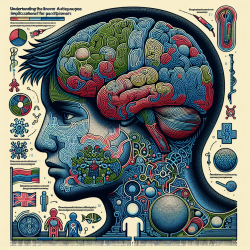
The world of Autism Spectrum Disorder (ASD) is complex and multifaceted, with each individual presenting a unique set of characteristics and challenges. As a practitioner working with individuals on the autism spectrum, staying informed about the latest research and technological advancements is crucial. One such advancement is the use of Magnetic Resonance Imaging (MRI) to study ASD. This blog will explore key findings from recent research and discuss how these insights can be applied in practice.
The Role of MRI in Autism Research
MRI has become an invaluable tool in understanding the structural and functional aspects of the brain in individuals with ASD. Two primary types of MRI are used in autism research: Structural MRI (sMRI) and Diffusion Tensor Imaging (DTI). These imaging techniques provide detailed insights into brain anatomy and connectivity, helping researchers identify potential biomarkers for early diagnosis.
Structural MRI: Mapping Brain Anatomy
sMRI focuses on capturing detailed images of brain structures. Research has shown that individuals with ASD often exhibit differences in brain volume and structure compared to neurotypical individuals. Notably, studies have observed variations in the amygdalae, cerebrum, and cerebellum across different life stages.
- Infancy: Some studies suggest that brain enlargement occurs around 12 months of age, which could serve as an early indicator of ASD.
- Childhood: Enlargements in cerebral gray matter (GM) and white matter (WM) have been noted, particularly in the frontal and temporal lobes.
- Adolescence and Adulthood: While some structural differences persist into adulthood, others may normalize over time.
Diffusion Tensor Imaging: Exploring Brain Connectivity
DTI provides insights into the brain's white matter tracts, which are crucial for understanding connectivity between different brain regions. Research using DTI has highlighted several key findings:
- Decreased Fractional Anisotropy (FA): Many studies report lower FA values in individuals with ASD, indicating potential disruptions in white matter integrity.
- Affected White Matter Tracts: Commonly affected tracts include the uncinate fasciculus, arcuate fasciculus, and cingulum bundle.
- Age-Related Changes: The impact of ASD on white matter appears to vary with age, underscoring the importance of longitudinal studies.
Implications for Practitioners
The findings from MRI studies offer valuable insights that can enhance diagnostic practices and intervention strategies for ASD. Here are some ways practitioners can apply these insights:
- Early Diagnosis: Understanding early brain changes can help identify children at risk for ASD sooner, allowing for earlier intervention.
- Tailored Interventions: Knowledge of specific brain structure variations can inform personalized treatment plans that address individual needs.
- Lifelong Monitoring: Recognizing that brain changes may continue into adulthood highlights the need for ongoing support and adaptation of interventions over time.
The Path Forward: Encouraging Further Research
The journey to fully understanding ASD is ongoing. Continued research using advanced imaging techniques like sMRI and DTI is essential to uncover more about this complex disorder. Practitioners are encouraged to stay engaged with current research and consider participating in studies that contribute to this growing body of knowledge.
If you're interested in delving deeper into this topic, I recommend reading the original research paper titled "Studying Autism Spectrum Disorder with Structural and Diffusion Magnetic Resonance Imaging: A Survey". This comprehensive survey offers a detailed overview of recent advancements in MRI applications for studying ASD.


