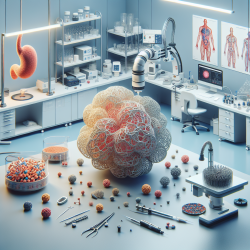
The field of cancer treatment is continually evolving, with researchers striving to develop more effective and less invasive methods. A recent study titled 3D-Printed Replica and Porcine Explants for Pre-Clinical Optimization of Endoscopic Tumor Treatment by Magnetic Targeting offers groundbreaking insights into the use of 3D printing technology for optimizing tumor treatments. This blog explores the study's findings and discusses how practitioners can implement these innovations to enhance their skills and improve patient outcomes.
The Promise of 3D Printing in Cancer Treatment
The study highlights the limitations of traditional animal models in cancer research, particularly when addressing anatomy-specific questions. The researchers propose using three-dimensional (3D) printed human replicas as a solution to these challenges. By creating patient-specific models from computer tomography (CT) data, they were able to fabricate anatomically accurate replicas that mirror human anatomy more closely than animal models.
This approach not only aligns with the "3R" principle—reducing, refining, and replacing animal experiments—but also provides a cost-effective and accessible method for preclinical evaluations. The use of open access software makes this technology available to a wide range of practitioners, even those with limited experience in 3D printing.
Practical Applications for Practitioners
The study's authors developed an eight-step process to create 3D replicas, which includes image segmentation, mesh refinement, and 3D printing. This method was successfully applied to fabricate a replica of locally advanced pancreatic cancer. Practitioners can use these replicas to practice endoscopic procedures and optimize the placement of magnetic field traps used in magnetic nanoparticle therapy.
The ability to accurately position magnetic field traps is crucial for effective tumor treatment. By practicing on 3D replicas, practitioners can refine their techniques and improve the precision of their interventions. Additionally, the study describes methods for stabilizing magnetic field traps using endoscopic clips, providing further opportunities for skill development.
Encouraging Further Research
The potential applications of 3D printing in cancer treatment are vast. While this study focuses on pancreatic cancer, the methodology can be adapted for other types of tumors and medical conditions. Practitioners are encouraged to explore how 3D printing can be integrated into their research and clinical practices.
Future research could investigate the use of different materials to better mimic the elasticity and mechanical properties of human tissues. Additionally, advancements in artificial intelligence could streamline the process of preparing CT data for 3D printing, making this technology even more accessible.
Conclusion
The integration of 3D printing into cancer treatment represents a significant advancement in medical research and practice. By adopting these innovative methods, practitioners can enhance their skills, improve patient outcomes, and contribute to the ongoing evolution of cancer treatment.
To read the original research paper, please follow this link: 3D-Printed Replica and Porcine Explants for Pre-Clinical Optimization of Endoscopic Tumor Treatment by Magnetic Targeting.


