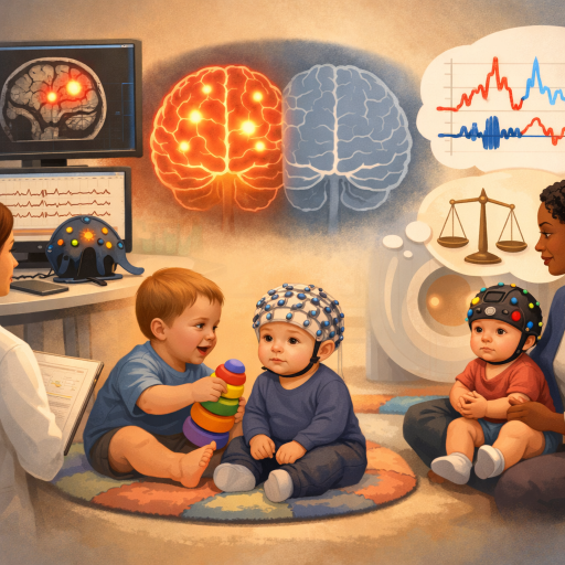
If you work with families who have an older child with autism spectrum disorder (ASD), you have probably fielded some version of this question from an anxious parent: Can you just scan the baby's brain and tell us now? The honest answer is no — but a set of six studies using EEG, fNIRS, structural MRI and home-video coding is building a genuinely useful clinical picture, as long as you keep straight what each study actually measured.
Only three of these six studies are prospective infant-sibling designs — the kind that follow high-risk infants forward in time before anyone knows which ones will receive a diagnosis. Study 1 tested 8-month-olds with an older sibling with ASD and followed them to age 3. Study 2 tested infants at 4–6 months and followed them to age 3. Study 5 scanned infants at 4–6 months and followed them to 36 months. The other three studies are not infant-sibling designs at all: Study 3 coded home video, ages 0–18 months, of children who were subsequently diagnosed with ASD, with the video coded retrospectively after diagnosis, compared with typically developing infants — there was no sibling-risk design and no later diagnosis to predict. Study 4 tested 3-year-olds already diagnosed with ASD against typically developing peers. Study 6 tested preschool-aged children already diagnosed with ASD against typically developing peers. So the age window this evidence actually covers runs from 4 months to preschool, not a single "first six months," and half of it describes children who already carry a diagnosis rather than infants whose future diagnosis is unknown at the time of testing.
That distinction matters for how you talk to parents and colleagues. Across these six studies, no single infant or cohort shows brain responses that are simultaneously too strong and too weak. What you see instead, when you line the studies up, is that different circuits — tested at different ages, with different tools, in different samples — sometimes look more reactive than typical and sometimes look less reactive than typical. That's a real and clinically relevant pattern, but it's a pattern assembled across separate groups of children, not a single dysregulated-in-both-directions signature inside one infant's brain.
Where the response looks "louder" than typical
Two of the six studies found evidence of heightened cortical response. In a 2019 study, 8-month-old infants with (n=116) and without (n=27) an older sibling with ASD were tested on a non-linguistic auditory oddball task. Relative to high-risk infants who went on to develop typically (n=44), the 14 infants who were later diagnosed with ASD showed reduced repetition suppression of gamma-band activity and significantly greater phase-locking to repeated tones — a pattern the authors describe as consistent with cortical hyper-reactivity and disturbed excitation/inhibition balance (Increased cortical reactivity to repeated tones at 8 months in infants with later ASD). Across the full high-risk sample, the size of this cortical reactivity index was dimensionally associated with slower language growth from 8 months to 3 years and with more severe parent-rated social communication symptoms at age 3.
A very different study, using a very different sample, found a related pattern at a much later age. A 2024 fNIRS study of preschool-aged children with and without ASD watched social versus nonsocial video clips. Children with ASD showed significantly higher medial prefrontal cortex activation specifically to social video stimuli, and this heightened activation correlated with more ASD symptoms on clinical measures (Prefrontal Cortex Responses to Social Video Stimuli in Young Children with and without Autism Spectrum Disorder). It's worth being precise about what these two studies share and don't: both found that more brain response to a stimulus category tracked with worse outcomes, which runs against a simple "less activity, less social processing" model. But they used different paradigms (auditory tones versus social video), different measurement tools (EEG versus fNIRS), different ages (8 months versus preschool), and different outcome measures — one is a prospective infant-sibling cohort, the other compares children who already carry an ASD diagnosis. They are not two runs of the same experiment; they're two independent hints pointing in a similar direction.
Where the response looks "quieter" than typical
Set those findings against a study that found the reverse pattern in early infancy. A 2017 fNIRS study followed infants at 4–6 months of age and measured brain responses to social videos (people playing peek-a-boo) versus non-social images (vehicles), and to human vocalizations versus non-vocal sounds. The 5 infants who were later diagnosed with ASD by age 3 showed reduced activation to visual social stimuli across inferior frontal and posterior temporal regions, compared with 16 low-risk infants, and reduced activation to vocal sounds paired with enhanced activation to non-vocal sounds in temporal regions, compared with both the low-risk group and 15 high-risk infants who did not go on to develop ASD (Cortical responses before 6 months of life associate with later autism). The degree of this atypical activation to both the visual and auditory stimuli correlated with parent-reported ASD symptoms in toddlerhood — the abstract doesn't specify which direction that correlation ran, so it's more accurate to say the degree of atypical activation tracked later symptoms than to claim less activation specifically predicted worse outcomes.
A 2016 home-video coding study found a behavioral echo of reduced responsiveness, though in a different design entirely: this one compared 10 children who were subsequently diagnosed with ASD against 10 typically developing peers, using home video from 0–18 months coded retrospectively after diagnosis, rather than following undiagnosed high-risk siblings forward in time. Analyzing 142 video sequences, researchers found a lower rate of vocalizations in the ASD group between 6–12 months, and fewer first words by 12–18 months, even though non-social babbling was actually more prevalent in the ASD group (Pre-linguistic Vocal Trajectories at 6–18 Months of Age As Early Markers of Autism). Note that these group differences emerged at 6–12 months and 12–18 months, not in the 0–6 month window, and the abstract does not report any analysis linking vocal patterns to later symptom severity. It's a small sample and home-video coding rather than lab-controlled measurement, but it's a rare direct look at real caregiver-infant interaction rather than a screen-based paradigm.
A third pattern: neither simply louder nor quieter
Not every study fits a hyper/hypo frame at all. A 2018 EEG and eye-tracking study of 3-year-old toddlers already diagnosed with ASD, watching dynamic social scenes, found frequency-specific alterations — in the theta and alpha bands — in the directed functional connectivity both driving and connecting from key social-brain hub regions (Early alterations of social brain networks in young children with autism). These were bidirectional, frequency-specific changes in network connectivity, not a straightforward reduction in activity. The most clinically interesting part of this study runs opposite to a "bigger deviation, worse outcome" story: within the ASD group, children who showed stronger recruitment of dorsomedial frontal, inferior temporal, and insular regions — activity the authors interpret as compensatory — had less atypical gaze patterns and lower clinical impairment. In other words, in this study, stronger activity in these regions was linked to a better outcome, not a worse one — a reminder that more brain activity is not automatically worse, and that some differences from the typical pattern may reflect the brain compensating rather than failing.
The contradiction you need to sit with
Put side by side, these six studies do not agree on direction, on age, on population, or on what a given deviation predicts. Auditory cortex at 8 months and medial prefrontal cortex in preschoolers: heightened response. Visual social regions and vocal responsiveness at 4–6 months: reduced response. Social-brain network connectivity in 3-year-olds: altered in a way that isn't simply more or less, and where more atypical activity in one specific pathway predicted a better outcome. This is not sloppy science; it likely reflects real differences in age, modality, which specific circuit was probed, and whether the children studied already carried a diagnosis or were still years away from one. The honest clinical takeaway is that there is no single "autism brain signature," even within this small literature. There are multiple, region-, age- and modality-specific findings, and while several point toward the same broad idea — that circuits involved in social and auditory processing look different early on — they don't all point the same direction, and they don't all speak to the same age window.
The one thing that doesn't quite generalize
It's tempting to draw a single unifying rule from all this: that the size of a deviation from typical predicts how a child does later, regardless of which direction that deviation runs. The evidence for that rule is thinner than it first looks. Study 1 found that a higher cortical reactivity index — more reactivity, specifically, not deviation in either direction — was associated with slower language growth and more severe symptoms, which is a directional finding rather than a magnitude-regardless-of-direction one. Study 2 found that the degree of activation to visual and auditory stimuli correlated with later parent-reported symptoms, but its abstract does not specify which direction that correlation ran, so it can't be used to confirm or contradict a directional account.
The rest of the studies don't support a shared magnitude rule either. Study 5's brain-behavior link is also directional: larger cerebellar and subcortical volumes were linked to more repetitive behavior, not simply "more different from typical" volumes generally. Study 6's link is directional in the same way: higher medial prefrontal activation, specifically, tracked with more symptoms. Study 4's brain-behavior finding runs the opposite way from a magnitude account: more compensatory activity was linked to lower impairment, not higher. And Study 3's abstract does not report any analysis linking its behavioral measures to symptom severity at all. So there is no single unifying law here, magnitude-based or otherwise, that this evidence supports — each study's brain-behavior finding has to be read on its own terms, for its own circuit, at its own age.
A structural clue that shows up before any task is given
One study didn't rely on a stimulus-response paradigm at all. A 2019 structural MRI study measured regional brain volumes in 4–6-month-old infants with (n=24) and without (n=26) an older sibling with ASD. This is a familial-risk comparison, not an outcome comparison: the volume difference was found between high-risk and low-risk infants, regardless of which high-risk infants later received a diagnosis. High-risk infants had significantly larger cerebellar and subcortical volumes than low-risk infants at this early age. Separately, and this part is outcome-linked, within the high-risk group, infants with larger volumes at 4–6 months went on to show more repetitive behaviors at 36 months (Familial risk of autism alters subcortical and cerebellar brain anatomy in infants and predicts the emergence of repetitive behaviors in early childhood). The authors themselves flag this as preliminary and in need of replication in larger longitudinal samples. It's a structural difference detectable before 6 months that predicted a specific behavioral domain three years later — but note that the initial group difference reflects family risk status, not eventual diagnosis.
What this evidence does not mean yet
These are small, group-level research findings, and the samples vary a lot in size and composition: Study 1's later-diagnosed group was 14 infants out of a 116-infant high-risk cohort; Study 2's later-diagnosed group was 5 infants, compared against 16 low-risk and 15 high-risk-without-ASD infants; Study 3 compared 10 already-diagnosed children against 10 typically developing peers; Study 5 compared 24 high-risk against 26 low-risk infants. Studies 4 and 6 don't report sample sizes in the material available. None of these studies used equipment or analysis pipelines available in a typical clinic, let alone a pediatrician's office, and none can currently tell you, for an individual infant, whether that child will receive an ASD diagnosis. Do not tell a parent that a brain scan can diagnose autism in infancy — it can't, not with today's evidence or today's tools.
What it does mean for practice now
What this evidence does support is a lower threshold for close behavioral monitoring in infant siblings of children with ASD, starting well before the 12-month mark that most screening timelines default to. Two of these studies — the ones that tested infants at familial risk and tied brain measures to eventual diagnosis — found measurable brain differences by 4–8 months: Study 1 at 8 months and Study 2 at 4–6 months. A third, Study 5, found a brain difference at 4–6 months that tracks familial risk rather than eventual diagnosis, alongside a separate within-group link between larger volumes and more repetitive behavior at 36 months. That's well ahead of when standardized screening tools like the M-CHAT are typically administered, and it's a defensible, evidence-based reason to advocate for earlier behavioral observation in siblings of diagnosed children, even though you can't order a scan to settle the question.
Worth being specific about what to watch for, and where the evidence does and doesn't reach. The domains with some infant-level support from this set are vocalization patterns and rate (Study 3, though its group differences appeared at 6–12 months and later, not before 6 months) and response to social visual and auditory stimuli during structured observation (Studies 2 and 5, at 4–6 months). Response to name, while a standard and reasonable item on any clinical surveillance checklist, isn't something any of these six studies measured — it's worth including in routine monitoring on general clinical grounds, not because this evidence base supports it specifically.
When you talk to parents, name both halves of the finding honestly: yes, researchers are finding measurable brain and behavioral differences in high-risk infants before their first birthday, in a small number of prospective studies. No, none of this translates into a clinical brain-based test today, and the studies themselves don't agree on a single direction of "different." What it does translate into is a rationale for watching more closely, starting sooner, and taking subtle behavioral signs in a sibling of a diagnosed child seriously rather than defaulting to "let's wait and see."
A therapist's early-monitoring checklist for high-risk infants
- Rate and quality of vocalizations during caregiver interaction, not just presence or absence of babbling — tracked longitudinally rather than at a single visit, since group differences in the available evidence emerged over months, not at one time point
- Attention to social video (as in peek-a-boo interactions) versus non-social visual stimuli during structured or naturalistic observation — the tested paradigm used social video and vehicle images, not isolated faces
- Response to human voice versus non-vocal sound during play
- Motor patterns and repetitive movements, tracked over time — a preliminary, unreplicated finding in one small high-risk cohort (n=24) linked larger cerebellar and subcortical volumes at 4–6 months to more repetitive behavior at 36 months within that group
- Growth trajectory of language and social communication milestones between 8 months and 3 years, since the strongest infant-level findings in this set used trajectory — not a single snapshot — as the outcome that correlated with early brain measures
None of these observations diagnose anything on their own. The prospective infant-level evidence behind them is thin — 14 later-diagnosed infants in one study, 5 in another, and a familial-risk (not diagnosis-based) comparison in a third — so treat these as reasons to document and refer earlier for a family that already has one child with ASD, not as a validated screening protocol. The direction of travel across Studies 1, 2, 3 and 5 — the ones that actually speak to the window before diagnosis or standardized screening — is that early brain and behavioral differences in autism are real and measurable in research settings well before standardized screening typically begins; what they are not, yet, is a substitute for careful behavioral observation over time.